
Clinical Photos


This is a root canal treated tooth that Dr. Keith extracted along with a large cyst full of bacteria. After she extracted the tooth, she used PRF (platelet rich fibrin) in the area to encourage healing.
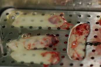
This is what the PRF (platelet rich fibrin) looks like just before it is placed. First, blood is drawn from the patient's arm, then is spun down in the centrifuge where all of the white blood cells and growth factors come together to form a clot as shown in the picture. During surgeries, Dr. Keith cleans the surgery site with ozinated gas and water to ensure there is no microscopic bacteria lingering, then places the PRF clot in the site before closing to optimize healing and aid in pain management.
This is a photo of an x-ray of a root canal treated tooth. The root canal tooth became reinfected and started to erode the bone surrounding the tooth.
This is a root canal treated tooth that Dr. Keith extracted for a patient. During a root canal, the nerve and blood supply to the tooth is removed and filled in with a rubber like material called gutta percha. Once the blood supply,vital nutrients and the body's immune system, are removed, the tooth becomes brittle and can easily break apart as shown in this photo.
This is another root canal treated tooth that became reinfected and created a cyst full of bacteria that began to affect the bone surrounding the tooth. Dr. Keith extracted the tooth and cyst and used PRF before closing.